What is Trichinellosis?

Trichinellosis, also known as trichinosis or trichiniasis, represents a parasitic zoonotic disease caused by the parasitic nematodes of the genus Trichinella. Human infection with this parasite stems from the consumption of raw (or semi-raw) meat of infected animals.
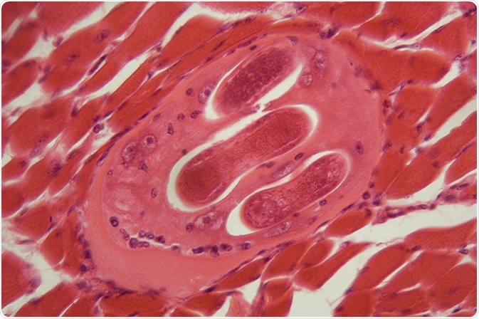
Credit: ChWeiss/ Shutterstock.com
The recognition of trichinellosis can be traced back to antique times, as historical references to similar clinical syndromes have been found in Egypt as early as 1200 BC. However, the modern scientific study of the parasite can be pinpointed to date from 1835, when physicians James Paget and Richard Owen from London observed parasitic larva in a human diaphragm.
This was followed by the provision of clear evidence of parasite transmission from animals to humans by Friedrich Zenker in 1860.
Today, trichinellosis and other parasites of the genus Trichinella attract the attention of a wide array of specialists: not only medical doctors, but also veterinarians, epidemiologists, biologists, biochemists, immunopathologists and geneticists.
Taxonomy and the diversity of species
Trichinella is classified under the phylum Nematoda, class Adenophorea, order Trichinellida and super family Trichinelloidea. There are two main clades that are recognized in the genus Trichinella – one containing species that encapsulate in the host tissue, and the second one that do not undergo encapsulation after they differentiate in muscle cells.
In addition to the best studied species known as Trichinella spiralis (which has a global distribution in a myriad of carnivorous and omnivorous animals), other species have been added to the classification. These include Trichinella pseudospiralis (which infests birds and mammals worldwide), Trichinella nelsoni (in African scavengers and predators), Trichinella nativa (in Arctic bears), and Trichinella britovi (in European and Asian carnivores).
Nematode morphology
Trichinella nematodes are 1-7 millimeters long and characterized by tapered and cylindrical anterior and posterior ends. Their elongated esophagus contains prominent stichocytes – i.e. a group of glandular cells that are arranged in a row along the posterior part of the organ.
Female worms are approximately twice as long as male worms. They have a single uterus that is full of developing eggs posteriorly, whereas the anterior part harbors fully developed juvenile worms. The females are viviparous, which means they lay live parasitic larvae.
Life cycle of the parasite
All species of Trichinella are characterized by a direct life cycle that completes development in a single host. The host capsule surrounding the infective larvae is a structure that stems from a modified striated muscle cell known as the nurse cell – a unique representation of true parasitism in the natural world.
This nurse cell is digested in the host’s stomach following ingestion of the infected muscle, which is followed by the movement of free larvae into the upper small intestine to invade the columnar epithelial cells. Within 30 hours the larvae undergo four molts to develop into mature male and female worms. Approximately five days after infection, the fertilized female worm begins to shed live newborn larvae.
The presence of adult worms in the human intestine may continue for weeks, during which time the emerging newborn larvae travel throughout the body via the blood and the lymphatic system. Although these larvae try to invade a plethora of different tissues, they can only mature if they successfully enter striated muscle cells.
Once they establish nurse cells in the muscle tissue, the parasite larvae can survive as such for years before calcification occurs. Such a hypobiotic stage can be maintained until the encysted larvae undergo ingestion by a new host. This is how humans get accidentally infected with Trichinella—when they consume improperly prepared meat of carnivorous animals, or eat food that is contaminated with such meat.
Sources
- http://cmr.asm.org/content/22/1/127.full
- https://www.ncbi.nlm.nih.gov/pubmed/17134656
- https://www.cdc.gov/parasites/trichinellosis/biology.html
- web.stanford.edu/group/parasites/ParaSites2005/Trichinella/trich.html
- vphcap.vet.cmu.ac.th/…/Chapter2.pdf
- Murrell KD. Helminthic Diseases: Trichinellosis and Zoonotic Helminthic Infections. In: Hamer D, Griffiths J, Maguire JH, Heggenhougen K, Quah SR, editors. Public Health and Infectious Diseases. Elsevier, 2010; pp. 327-332.
Further Reading
- All Trichinellosis Content
- Trichinellosis Diagnosis
- Trichinellosis Epidemiology
- Trichinellosis Symptoms
- Trichinellosis Treatment and Prevention
Last Updated: Feb 27, 2019

Written by
Dr. Tomislav Meštrović
Dr. Tomislav Meštrović is a medical doctor (MD) with a Ph.D. in biomedical and health sciences, specialist in the field of clinical microbiology, and an Assistant Professor at Croatia's youngest university – University North. In addition to his interest in clinical, research and lecturing activities, his immense passion for medical writing and scientific communication goes back to his student days. He enjoys contributing back to the community. In his spare time, Tomislav is a movie buff and an avid traveler.
Source: Read Full Article